Metabolic Imaging Core
The Metabolic Imaging Core links a variety of imaging facilities providing services to NORCH investigators who currently require or plan to conduct human studies using state-of-the-art imaging research instrumentation and expertise. Our goal is to provide a central access point for investigators to conduct human studies that include imaging technology for in vivo phenotyping of human tissues for nutrition, obesity, and metabolism studies.
Getting started:
Interested in utilizing the services and equipment of the Metabolic Imaging Core at the NORCH? Hear from Dr. Martin Torriani on how to get started.
Core Services
Individual consultation and training services:
-
Imaging analysis, including quantitative interpretation of imaging data
-
H-MRS for lipid profiling (intramyocellular lipid, intrahepatic lipid, marrow fat)
-
Help with study design and methods to include available Core techniques in human research studies.
-
Tutorials and workshops in imaging techniques used in metabolic investigation
MRI and MRS Services:
-
Quantification of areal and volumetric visceral and subcutaneous adipose tissue
-
Liver MRI for assessment of steatosis and fibrosis (using Perspectum Multiscan protocol)
-
MR elastography (in development) for assessment of liver fibrosis
-
Brain MRI
-
Functional MRI
-
Additional custom MRI protocols available for NORCH-related research
-
MRS of muscle, liver, marrow and adipose tissue with analyses for lipid profiling
CT, PET, and Other Services
-
Quantitative CT (helical) using single- or dual-energy CT for volumetric acquisition of bone mineral density
-
Quantitative CT (single-slice) for quantification of visceral and subcutaneous fat, cross-sectional muscle area
-
Ultrasound and shear-wave sonoelastography for assessment of liver fibrosis and steatosis
-
CT analysis for body composition performed with deep learning algorithm (ideal for medium or large-scale studies or datasets requiring short-term execution)
-
Image-guided biopsies
-
PET, PET/CT, and PET/MRI
-
Other services may be available upon request
Equipment
Click for the full list of equipment
- Perkin Elmer fluorescence spectrometer
- Inverted and epi fluorescence Zeiss microscopes
- Nikon Eclipse fluorescence microscope
- Union Biometrica COPAS BioSort robot
- Titertek MapC2 Liquid and Agar Dispenser
- Illumina HiSeq 2500
- Illumina NextSeq 2000
- Illumina MiSeq
- Illumina CBot
- 10 x Genomics Chromium
- Covaris S2 shearing device
- Agilent BioAnalyzer
- BioRad qPCR
- Flow cytometers (Attune NxT Flow Cytometer)
- Vario-MACS immunomagnetic cell sorting unit
- Beckman-Coulter DU-640 spectrophotometer
- Neon Transfection System
- ChemiDocMP Imaging System
- SpectraMax ABS Microplate Readers
- Luminex MAGPIX System
Inquiries and Scheduling
Office Hours: Fridays at 9am
Dr. Torriani holds office hours on a weekly basis for questions related to the services offered above. Zoom details can be requested via the inquiry form.
Interested in utilizing our services or equipment for a current or upcoming project? Fill out the inquiry form below.
Core Leadership
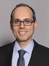
Martin Torriani, MD
Primary Contact
Director of Metabolic Imaging Core
Professor of Radiology
mtorriani@mgh.harvard.edu
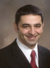
Georges El Fakhri, PhD
Co-Director of Metabolic Imaging Core
Professor of Radiology
gelfakhri@mgh.harvard.edu

Bruce Rosen, MD, PhD
Laurence Lamson Robbins Professor of Radiology
Director, Athinoula A. Martinos Center for Biomedical Imaging